 A research team led by Prof. HENG Pheng Ann, Department of Computer Science and Engineering has developed an automated image processing technology that, through deep learning, is able to offer efficient and accurate diagnosis using CT scan and histopathological images. The technology has been tested on two of Hong Kong’s most prevalent cancers — lung cancer and breast cancer, achieving diagnostic accuracies of 91% and 99 percent respectively in durations of between 30 seconds and ten minutes. The tests demonstrate that the technology not only boosts efficiency in clinical diagnosis, but also reduces misdiagnosis. The automated screening and analysis technology is expected to be widely adopted by the local medical sector in the next couple of years.
Detection of pulmonary nodules through Deep Learning
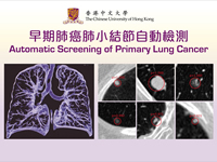
Lung cancer has been the leading cause of cancer death in Hong Kong. At an early stage, lung cancer mostly exists in the form of small pulmonary nodules, which appear on medical images as shades of small lumps. Currently, doctors depend on chest CT scans to reveal those nodules. However, each scan often results in hundreds of images. Assuming that going through each image requires 3 seconds, an analysis of these images by the naked eye will take 5 minutes to complete. Such examinations are time consuming, and must rely on the doctors’ experience and sharpness of focus. When Prof. Heng and his team apply deep learning technology to CT scans, they are able to locate the pulmonary nodules in 30 seconds, with an accuracy of 90%.
 The technology, which the CUHK team started working on five years ago, is at the forefront of international medical technology. With positive feedback from the medical sector, Professor Heng expects it to be widely adopted in the next couple of years. ‘Deep learning makes use of advanced training to improve the sensitivity of the technology, so that it is able to tackle a major challenge that a naked-eye examination faces – that is, removing noise and reducing false positives,’ said Professor Heng. He went on to disclose that, in order to further improve the technology, the team would be working with top hospitals in Beijing, to provide solid evidence in support of early diagnosis and treatment of lung cancer.
Automated Detection of Metastatic Breast Cancer in Histology Images
Since 1990, the number of breast cancer patients in Hong Kong has been consistently on the rise. It is the most prevalent cancer amongst local women, and the third amongst all cancers. To determine whether a patient has the cancer, doctors often must extract and examine live tissue samples. Using mammograms or MR scans to locate the lump, samples are extracted and examined under the microscope to see if there are signs of tumour and whether the tumour is benign or malignant. A digital histology is of high resolution, often up to one gigabyte in file size – equivalent to a 90-minute high resolution movie. Examining such an image requires a lot of time and energy.
To solve the problem, the CUHK team has developed a novel deep cascaded convolutional neural network to process the histopathological images. Making use of a fully convolutional network, the model can efficiently and accurately detect the metastatic cancer with a high-resolution score-map. The whole automated analysis process takes about 5~10 minutes, as compared to the 15~30 minutes that are required if examined by the naked eye. In terms of accuracy, the system has achieved a rate of 98.75%, 2% higher than analysis conducted by experienced doctors. This indicates that it is an invaluable reference for clinical diagnosis on breast cancer.
A key advantage of artificially intelligent deep learning is that it is able to analyse large quantaties of parameters. The more the data, the higher its accuracy. When this automated screening and analysis system is applied to the medical sector, it acts as a tireless assistant to the doctor that can quickly identify the source of an illness, enabling a timely and appropriate treatment.
In the News