Anterior Segment Analyzer (Pentacam)
A device measures and analyses various anterior segment parameters, including corneal wavefront analysis, corneal topography and 360 degree automatic chamber angle measurement. It provides information for the diagnosis and management of glaucoma, cataract surgery and LASIK surgery.
Anterior Segment Optical Coherence Tomographer
A device examines the cross-sectional imaging of the cornea, anterior chamber and the bleb segment of the sclera in three-dimensional view. It also measures curvature, length, area and volume of the anterior segments by computed analysis. It provides information for the diagnosis and management for wide variety of clinical situation such as patients with corneal anomalies and glaucoma.
Corneal Biomechanical Analyzer / Tonometer
A device measures the intra ocular pressure (IOP), corneal thickness, dynamic corneal response and biomechanical properties. It determines the influence of the corneal biomechanical properties on conventional IOP measurements and provides information for the diagnosis and management of glaucoma and LASIK patients.

Corneal Topography
A non-contact device measures the optical aberration of eye, topography of cornea, refractive error, corneal and pupil parameters. It provides information for the diagnosis and management of cataract, corneal diseases and planning before refractive surgery.
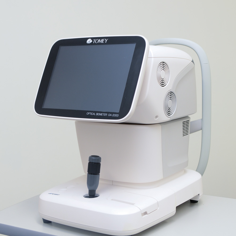
Optical Biometry
A non-contact optical biometry device measures the length of eyeball, depth of aqueous chamber and surface curvature of cornea. It provides information for measuring the power of intraocular lens before cataract surgery and monitors eyeball elongation in children regarding the myopia progression.
Optical Coherence Tomography Angiography
A device that provides angiography without needing intravenous dye injection detailed assessment of the retinal and choroidal vasculature. It provides information for the diagnosis and management for wide variety of clinical situation such as patients with glaucoma, diabetic retinopathy, macular degeneration and other retinal diseases.
Spectral-domain optical coherence tomography and Confocal Scanning Laser Ophthalmoscope
A device with both spectral-domain optical coherence tomography and confocal scanning laser ophthalmoscopy provides us the information of the nerves, vessels and macula integrity of the retina. Spectral-domain optical coherence tomography can access the morphology and quantitatively measure the details of retinal structure (e.g. macular thickness and retinal nerve fiber layer thickness) as well as comparison with a built-in normative database. Dye angiography with confocal scanning laser ophthalmoscope provides high-resolution images and video of the dynamic movement of blood flow and leakage through the retinal and choroidal vessels. It helps to illustrate the retinal and choroidal microvasculature for retinal diseases such as diabetic macular edema and age-related macular degeneration. The device provides information for the diagnosis and management for wide variety of clinical situation such as patients with glaucoma, macular anomalies and degeneration.
Ultra-widefield Colour Fundus Photography
A device provides non-mydriatic ultra-widefield retinal images, up to 200 degrees. It allows the examination of the periphery of the retina. It provides information for the diagnosis and management for wide variety of retinal diseases such as diabetic retinopathy and retinal vein occlusion.
Visual Field Test
An automated device that can detect dysfunction in central and peripheral vision. It provides information for the diagnosis, management and monitoring of glaucoma and optic nerve abnormalities.
| MON - FRI (Except on Public Holidays) |
| 0900 - 1800 |
| Telephone : (852) 3943 5886 Whatsapp : (852) 9681 4010 |
| Email : eyecentre@cuhk.edu.hk |
| 3/F, Hong Kong Eye Hospital, 147K Argyle Street, Kowloon, Hong Kong |


