News Centre
CUHK Develops Automatic Retinal Image Analysis Technology for Identifying AutismMaking Objective Screening and Early Intervention Possible
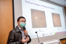
The Faculty of Medicine at The Chinese University of Hong Kong (CU Medicine) has successfully developed the Automatic Retinal Image Analysis (ARIA) technology to assess the risk of autism spectrum disorder (Autism). It is done by analysing the captured retinal images to identify if there are any retinal characteristics such as those related to thinning of the retinal nerve fibre layer of the children’s eyes. The research team hopes that ARIA technology can be used as a risk assessment tool for autism screening and provide early intervention instead of waiting for a lengthy diagnosis, so that a difference can be made through a long term positive impact on the life of autistic children. Results of the study can be found in the journal EClinicalMedicine published by The Lancet.
The lack of an objective screening method in young children is a major cause of delayed diagnosis or even misdiagnosis
Autism is a complex neurodevelopmental disorder that begins at a young age, and it affects many aspects of the individual’s life, including social interaction, communication and the way sensory information is being processed. Early intervention is highly recommended to achieve a better outcome. However, the diagnosis of autism requires assessment by a multi-disciplinary team of professionals mainly relying on the questionnaire and requires a lengthy assessment duration. The lack of an objective screening method, especially in young children is also a major cause of delayed diagnosis or even misdiagnosis.
Previous studies have shown that thinning of the retinal nerve fibre layer was significantly associated with high functioning autism spectrum disorder and Asperger syndrome. It is suggested that retinal images may provide critical information for the classification of the disease, and may contribute to the development of an objective measure of autism.
Study shows that ARIA technology can identify autistic patients with sensitivity up to 90%
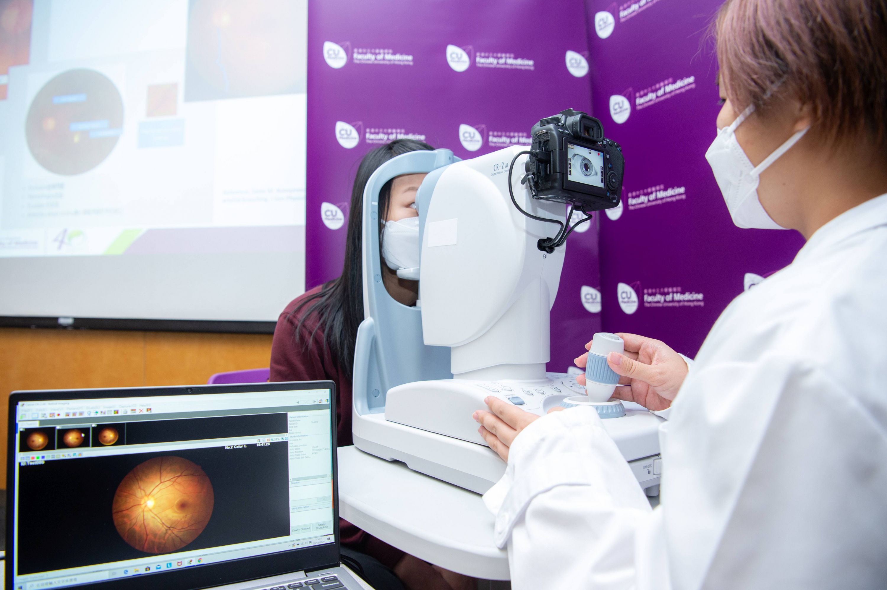
Because of this, the Centre for Clinical Research and Biostatistics of the Jockey Club School of Public Health and Primary Care has developed the Automatic Retinal Image Analysis (ARIA) technology to assess the risk of autism. The research team recruited 46 autism cases from special schools run by Hong Chi Association, followed by age-gender matching for 23 pairs of autism-control subjects. The team captured retinal images of all participants using a non-mydriatic fundus camera and used ARIA to optimise the information of the retina to develop a classification model for autism.
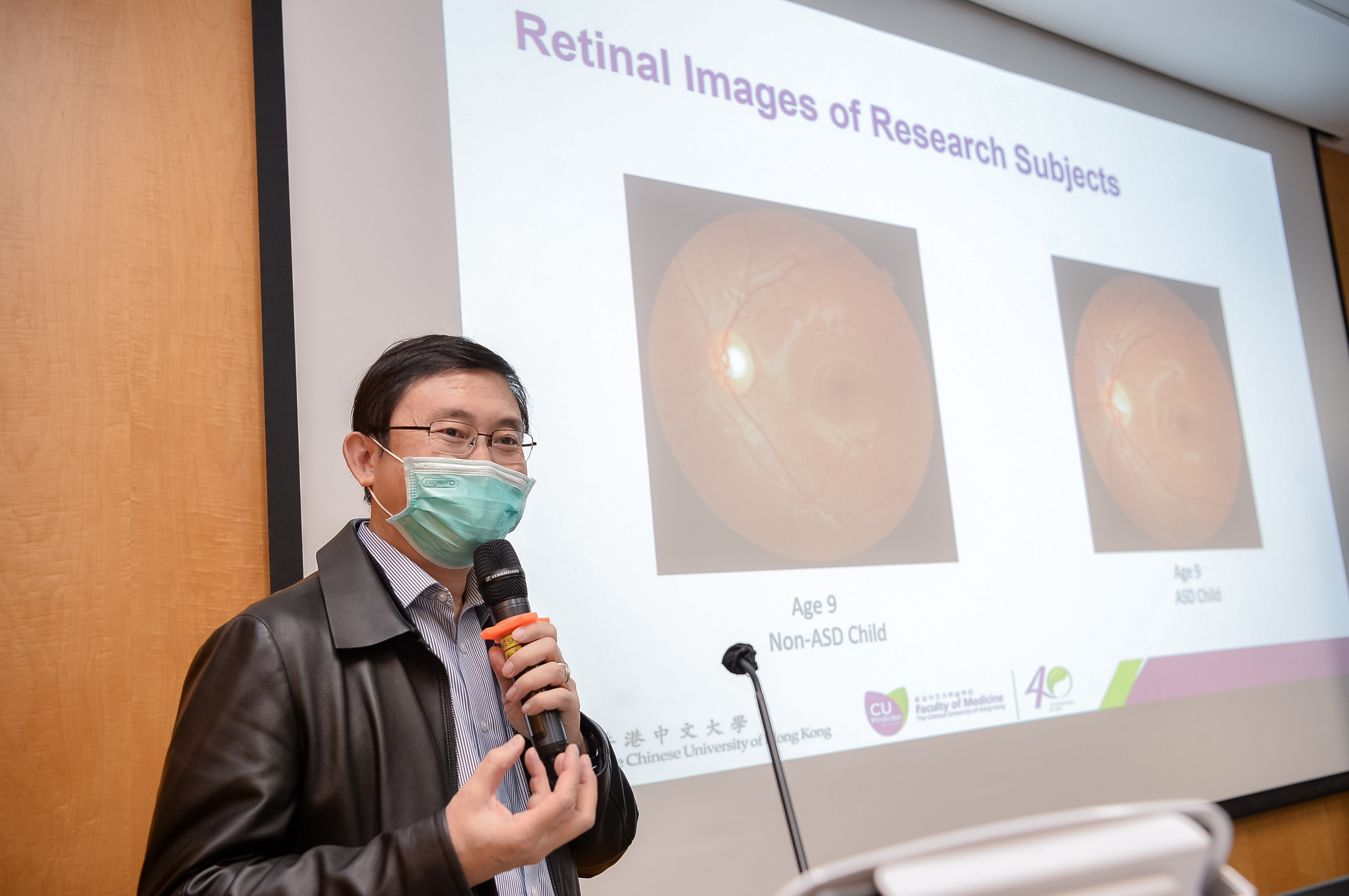
Study results show that autism subjects have significantly a larger optic disc diameter and larger optic cup diameter. The sensitivity and specificity of this technology to identify autism were 96% and 91% respectively.
Professor Benny Chung Ying ZEE, Director of the Centre for Clinical Research and Biostatistics, the Jockey Club School of Public Health and Primary Care (JCSPHPC) at CU Medicine, who led the study, remarked, “The use of retinal image analysis is non-invasive, fully automatic and relatively convenient. Retinal images can be obtained from very young children to assess their risk of suffering from autism. This technique provides an objective screening method that can be implemented in a community setting and provide an efficient tool to assess the risk before clinical and behavioural assessment.”
Closer collaboration with the community for deeper social impact
Ms Maria Ming Po LAI, Nursing Officer and Assistant Director of the Centre for Clinical Research and Biostatistics, JCSPHPC at CU Medicine, and first author of this publication, added that the importance of this study lies not only in the scientific and technological advancement, but also shows how the close collaboration between The Chinese University of Hong Kong, Schools of Hong Chi Association, and optometrists in the local community can improve social capital and realize the common goal of achieving social impact.
Ms Sally Chung Man CHIU, Educational Psychologist at Schools of Hong Chi Association, stated, “Every student’s need is unique, and the needs of students with autism and special educational needs (SEN) are even more diverse. We truly appreciate CU Medicine for inviting students of Hong Chi Association to take part in this research. Through developing such innovative medical technology, and with the partnership of optometrists in the local community, this research not only helps assess children’s risk of autism to identify appropriate services at an early age, but it also enhances public understanding about the needs of children with autism and SEN, thus bringing more social acceptance and inclusion to society.”
Research team receives the Outstanding Social Capital Partnership Award for the development of ARIA technology
ARIA technology has been applied to assess the risk of stroke and risk of white matter hyperintensities as an early risk factor of dementia. The research team has just received the Outstanding Social Capital Partnership Award and the Social Capital Builder Logo Award from the Community Investment and Inclusion Fund of the Labour and Welfare Bureau, Government of the Hong Kong Special Administrative Region, for their outstanding contributions to the development of social capital for health promotion in the community using ARIA technology.
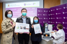
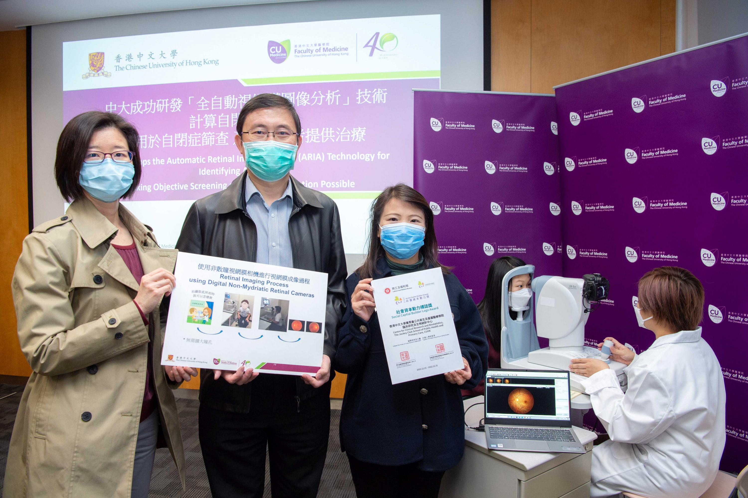
CU Medicine has successfully developed the Automatic Retinal Image Analysis (ARIA) technology to assess the risk of autism in children by analysing their captured retinal images, in order to identify if there are any retinal characteristics related to autism. The research team has just received the Outstanding Social Capital Partnership Award and the Social Capital Builder Logo Award from the HKSAR Government to recognize the contribution from the team to the development of social capital by the application of the ARIA technology. From right: Ms Maria LAI, Assistant Director; Prof. Benny ZEE, Director of the Centre for Clinical Research and Biostatistics of the Jockey Club School of Public Health and Primary Care at CU Medicine; and Ms Sally CHIU, Educational Psychologist at Schools of Hong Chi Association, which is the partner of this research project.
