Easy Screening for Skin Cancer

(Photo courtesy of Information Services Office, CUHK)
Terahertz imaging may enable professionals to soon find a faster, safer and more efficient way of treating skin cancer and other skin-related maladies.
Scientists from the Faculty of Engineering at CUHK and the University of Warwick have developed a novel method for analysing skin structure using a type of radiation known as Terahertz radiation (T-rays). Based on the research led by medical physicist Professor Emma MacPherson, this latest innovation may enable professionals to soon find a faster, safer and more efficient way of treating skin cancer and other skin-related maladies.
Skin Cancer Concerns
According to the World Health Organization, currently around 2-3 million non-melanoma skin cancers and 132,000 melanoma skin cancers occur globally each year. Skin cancer can be difficult to assess. Small changes to the skin’s surface can be part of a larger tumour with the cancer spread much farther than is visible. Currently, doctors removing a surface-level tumour must effectively take multiple biopsies to ensure they have reached the margin of the cancer and removed it all. This time-consuming process extends the surgery so that a simple procedure can take three hours or more while the tests are run.
T-rays Give a Clearer Picture
The research team aims to use Terahertz imaging to allow surgeons to map the cancer before surgery is started to get a better idea of how far a tumour below the skin surface has spread. Not only would this save much of a surgeon’s time during surgery, doctors could also prepare appropriate skin grafts and warn the patient of the likely extent and result of any surgery.

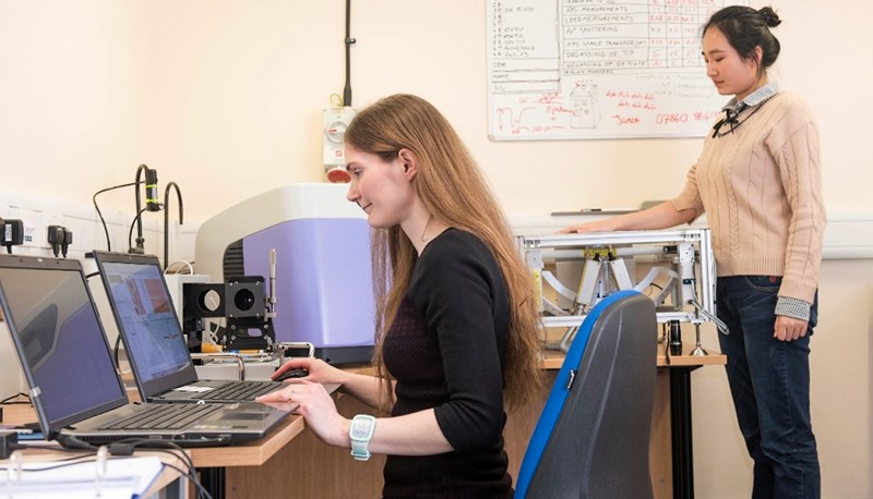
By measuring the Terahertz response of skin, the imaging system developed by Professor MacPherson (left) can assess the hydration and structure of that skin to identify skin cancer and other skin-related maladies.
Terahertz imaging is very sensitive to water concentrations. By measuring the Terahertz response of skin, the system can assess the hydration and structure of that skin. So how does the science work? T-rays sit in between infrared and Wi-Fi on the electromagnetic spectrum. The low energy photons of T-rays are non-ionising, making them safe in biological settings like medical screening. Professor MacPherson’s system emits lower background radiation than is emitted by a human body. The terahertz waves are created by shining a pulsed laser at a gallium arsenide semiconductor, which when excited emits the right frequency. Only the T-rays passing through the outer layers of skin before being reflected back can be detected. Depending on the properties of the skin, those T-rays will be reflected back slightly differently. Scientists can then compare the properties of the light before and after it enters the skin, gaining more detailed information.
Redefining Skin Treatments
Professor MacPherson has a concurrent post at the University of Warwick in the UK. She will work with University Hospital Coventry at Warwick to start testing the imaging in the clinic with patients. Her ultimate goal is to have a plugin terahertz device for a mobile phone so that people can run the test reliably at home.
The terahertz system could also be used for other skin treatments. For instance, to help improve the diagnosis and treatment of dry skin conditions such as eczema and psoriasis by building a more detailed picture of the structure of an area of skin. T-rays can detect minute changes to the hydration of skin, allowing for quantitative assessments of how the skin is improving with different products.
Read more: